Zooming in on a career looking through the lens
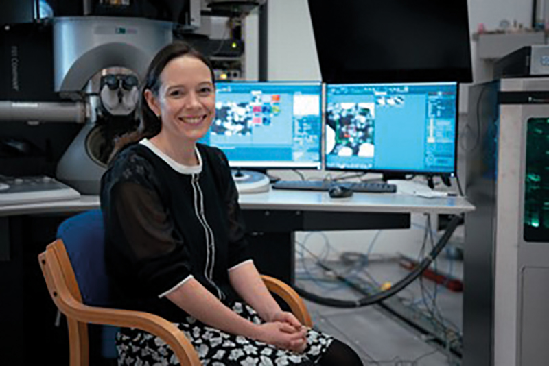
Sarah Haigh is at the helm of a project at the University of Manchester, UK, to develop a unique transmission electron microscope that integrates artificial intelligence and automation.
Perhaps Sarah Haigh was always supposed to be a microscopy specialist. Certainly, she had strong influences towards STEM early. Her mother was a Maths teacher and her father a systems engineer, having studied Maths at university.
Haigh hung onto Maths for her A-levels, as well as Physics and Chemistry – a classic science triumvirate. But choosing Art as well was a curveball.
It was 'an unusual combination, but actually turns out to make sense for microscopy', she reflects. Being a visual medium, there is an artistic element, 'although I don’t photoshop my images, let’s just be clear', Haigh points out.
Her reply to her headmaster of 'Because STEM’s better', when he questioned why she was choosing Materials Science over English at university, seems self-assured and clear-minded for a young woman who was somewhat of an all-rounder.
She notes that while studying Space is 'cool', she wanted to have a more immediate impact.
It was an open day at Oxford University on Materials Science that sparked her interest. Haigh asked her Chemistry teacher what it was about, to be told that he had no idea, and she should go and find out.
She is grateful for the impetus. 'I met a really inspiring technician who was running the undergraduate labs, and she was passionate about the role of Materials Science in saving the world – creating cars that don’t rust or more efficient batteries. And that was what inspired me to try and apply to Oxford and to other universities to study Materials Science.'
Admitting that she did not like learning organic chemistry formulas, Materials Science seemed to bring together more inorganic chemistry and parts of Physics about understanding how processes work.
A new perspective
The lab technician she met at the open day at Oxford continued to spark Haigh’s imagination while prepping equipment during her undergraduate studies – giving advice that was both relatable and practical.
During her subsequent Master’s research project on creating less expensive superconductors, she was privileged to use an expensive bit of kit called a nano secondary ion mass spectrometer (nanoSIMS), which can characterise different elements in materials at high spatial resolution with high sensitivity.
As she finished her project using the equipment, an even more expensive machine – a world-first transmission electron microscope (TEM) – was being installed just down the corridor from where she was working. She applied to the investigator who was leading that instrument, Professor Angus Kirkland, and subsequently moved over to use that for her PhD.
Although the microscope was purchased, it was a prototype system, so Haigh visited JEOL’s manufacturing headquarters in Akishima near Tokyo, Japan, on a few occasions. Her PhD was to effectively push the capability of the instrument to demonstrate images where white dots correspond to the positions of individual atomic columns. They were searching for ways to achieve better spatial resolution so as to see atoms closer together and get access to more materials.
She explains, 'Although first invented in 1931, electron microscopes were not initially better than optical ones. In 1998, a new generation of microscopes began to be sold, ones that had aberration correctors, which is effectively like putting a pair of spectacles on when your eyesight is a little bit short-sighted, or long-sighted. These are flexible spectacles, so they can vary depending on the errors that are present in the microscope on that day. The TEM instrument that I used in my PhD was the first to have not just one pair of spectacles but two, allowing it to operate in several different modes and still produce visuals of atoms.'
But 'one of the frustrations', Haigh shares, was that the advances in microscopy she achieved during her PhD only worked on model systems and did not work on more challenging materials problems like batteries or fuel-cell electrodes.
This fuelled her focus to solve Materials Science problems, rather than focussing on pushing the limits of instrumentation, or at least doing so with a particular application or problem statement in mind.
Haigh is unapologetic about reverting to her Materials Science roots. 'I think of myself as a microscopist trying to solve problems, rather than somebody who’s trying to develop a better microscope for the microscope’s sake,' she elaborates.
There are a 'few occasions in my career where I can point to something where we’ve had a tiny role in making something…that says new and improved on the label, and I think that’s a real buzz.'
Pet project
After her PhD, Haigh thought it would be fun to sell microscopes, and had flexible funding from Oxford to spend half her time working at the university and the other half working for JEOL.
She worked as an application specialist and put in the first aberration-corrected spectacle microscopes in South Africa, which was a 'fabulous experience'.
As there was a lack of advanced microscopy expertise country-wide, JEOL provided training, including at the Nelson Mandela University in Port Elizabeth. 'Being a part of that installation and development, and the pre-training before the system arrived, was a hugely privileged role to have had,' enthuses Haigh.
She was clearly always drawn to the newest shiny microscopes, reflecting, 'Are you noticing a trend yet?'
It was only a matter of time before Haigh needed her own shiny bit of kit. “Rather than continuing to install them for others, I had pet projects I wanted to have a play with. The only way that you really get the money for your own pet microscope is to be based in a university and running a research lab or a research centre.'
So, 18 months after her PhD, she applied and was accepted for a lectureship position in Materials at the University of Manchester, UK.
Initially, she found it 'absolutely terrifying' being an independent academic without the guidance of her PhD supervisor or boss at JEOL. Having not been 'a proper postdoc', Haigh felt very unprepared.
But unusually, Manchester appointed around 10 lecturers at the same time in different disciplines across the university, all with some relationship to graphene. 'That was a fantastically supportive and inspiring community to go into,' Haigh shares.
As many of her colleagues also stayed on and became professors, she still collaborates with them on various opportunities. 'Being around a group of smart people coming up with unusual problems is ideal for a microscopist – all I need to do is to convince them that the answer could come from microscopy.'
And she had her hands on some great equipment too, to seal the deal. Manchester has a large Electron Microscopy Centre and Haigh knew that the university had applied for an aberration-corrected microscope. That instrument arrived about 18 months after she started, but in the interim, some colleagues at Liverpool very kindly allowed her to use their one.
An alternative view
'I was really lucky that, just as they (Manchester University) appointed me, there was the whole graphene thing blowing up in 2010, and the Nobel Prize was awarded to its inventors based at Manchester. I did a lot of work in graphene. I had a crazy idea that I’d look at graphene from the side rather than from the top.'
The Manchester group was building transistor geometries by layering 2D graphene and hexagonal boron nitride on top of each other. By looking side-on, Haigh saw that the rippled effect of graphene determined how well it performed in the transistors. This has become one of her most cited works. She found graphene behaves better when that rippled effect can be 'ironed out'.
'It turns out, taking a slightly different view (literally) from other people yielded information that no one else had seen before, and it helped establish one of my research directions, which was cross-sectional imaging of different 2D materials. Understanding layer interactions, which dominates behaviour in lots of different areas, has galvanised this field.'
Shiny, happy, epic TEM
And her love affair with microscopes continues. 'I’ve had a few large grants in my career – EvoluTEM, SoluTEM and AutomaTEM – they all have TEM at the end, it is a very bad pun, and I apologise for it, Haigh wryly observes.
AutomaTEM – which is the name of the latest project – is next on the hit list. The Manchester team, led by Haigh, will be among the first scientists in the world receiving a state-of-the-art new type of scanning transmission electron microscope – an Iliad from Thermofisher Scientific, capable of atomic-scale elemental and chemical mapping in just a few seconds.
Haigh’s team combines this instrument with advanced computation to build new capabilities for automation and data analysis, for an instrument that can acquire huge datasets of local chemical information in days rather than years and with minimal human input.
Heralded as a world-first TEM, it will integrate artificial intelligence and is funded through a £9.5mln project supported by the Engineering and Physical Sciences Research Council (EPSRC), the University of Manchester, the Henry Royce Institute and bp, in collaboration with manufacturer ThermoFisher Scientific.
It is hoped the instrument will accelerate innovation in materials for quantum computing, low-power electronics and new catalysts to support the energy transition.
Haigh notes how cryo-electron microscopists currently turn up to analyse five samples and have completed this two days later with all their images and with minimal hands-on operation of the TEM – this is not what happens in materials.
Ideally, with the AutomaTEM, researchers will be able to load a sample and come back a few hours later to collect 10,000s of images showing the composition of all the nanoparticles or of all the grain boundaries in their sample.
'The elemental mapping is much faster than anything we’ve had before, about five times faster. So, the individual data collection will go from being a few minutes to less than a minute,' says an excited Haigh.
The team hopes to train the microscope on catalytic nanoparticles. She adds, 'It doesn’t matter if we miss 1% of the particles when we’re trying to get information about 100,000 particles, as long as we’re not systematically missing the same type of particle. If we don’t find them all, there isn’t an issue…We are not looking for the accuracy that you might need from machine learning in other areas. We’re just looking for it to be able to do it as well as a human but much more quickly.'
It is estimated that the new microscope will generate hundreds of terabytes everyday and the team has budgeted for instant access to the data for three months. The university has invested in servers to deliver this capability, absorbing learnings about data storage and dissemination protocol from the National X-ray CT Centre, also at the University.
Having to manually train the new microscope is the current rate limiter. This involves labelling images and telling the computer what it should be looking for.
But Haigh believes that this new materials characterisation capability will open up roles for more researchers not less, to analyse 'richer datasets and then develop improved materials based on that improved characterisation”.
She hopes it will become a widespread research tool to limit the time spent procuring data and represents 'democratisation of atomic-scale characterisation', so that you don’t need to be an expert microscopist to access such information on your sample.
Big picture
When we spoke in the summer, Haigh had just returned from the Royal Society Summer Science Exhibition 2024, hosting on one of the 14 main exhibits. Over the course of five days, she spoke to the general public and schools about developing new materials with applications in quantum computing, 'ad-libbing' a bit from her specialism in materials characterisation.
'One minute you are explaining your exhibit to an interested six-year-old and their parent, then a minute later you are being questioned by a Fellow of the Royal Society, so you have to be careful to judge your audience,' Haigh flags.
In the course of our conversation, she seems to be very well versed in pitching the right level of complexity and detail in her answers to my questions. She is expansive while sticking to the subject, and able to zoom out from too much detail – which seems a contrary skill to that of a microscopy specialist.
She disagrees, suggesting instead that 'microscopy is about trying to work out how the tiny piece of a science puzzle that I can see in my image, could make a wave which might change the macroscopic world around us for the better.'